Through detection of increased glucose metabolism, PET can also be used to detect active inflammation in the myocardium. This can occur, for example, in myocarditis, sarcoidosis, or in case of infection of a valve prostheses in endocarditis. It is important that the patients follow a strict diet without consuming carbohydrates the days before examination. Please consult the information leaflet.
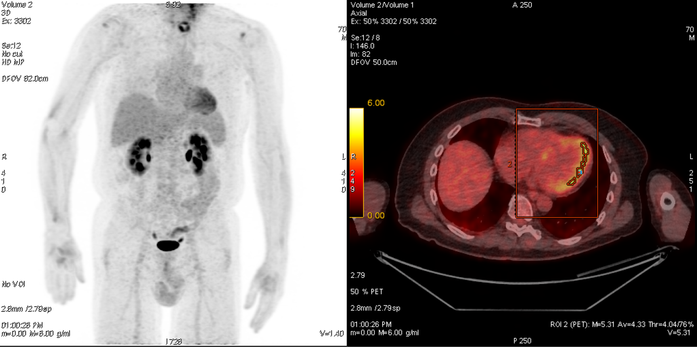
70-year-old patient with probable cardiac sarcoidosis, which could be clearly diagnosed by means of 18F-FDG PET / CT: The maximum intensity projection (left) shows clear metabolic activity in the left ventricular myocardium. The fused PET / CT (right) shows the increased activity (yellow) mainly in the lateral wall of the myocardium. In addition we can quantify the activity with PET (SUVmax here 5.31) which is particularly helpful for therapy monitoring.
In selected cases, especially when examining myocarditis or sarcoidosis, it can be helpful to combine PET and cardiac MR, available at our clinic in Schlieren. This combination leads to increased diagnostic accuracy and the spectrum of clinical questions to be addressed is increased.
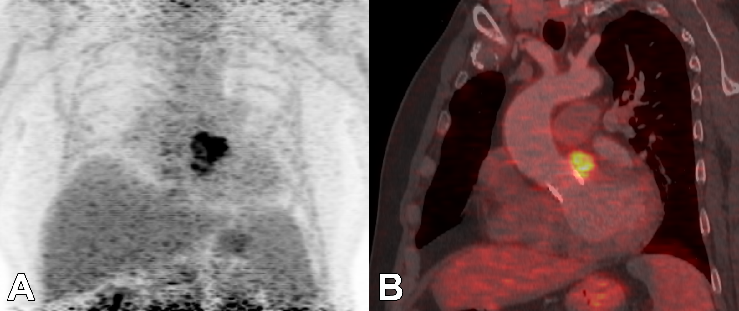
63-year-old patient after biological aortic valve replacement. Echocardiographic with probable paravalvular abscess that can be clearly diagnosed using 18F-FDG PET / CT. Maximum intensity projection (MIP) with clear focal metabolic activity projected onto the aortic root (A), which can be clearly localized in the series fused with the CT (B).