We provide a state-of-the-art MR and a combined PET / MR service for cardiac imaging in our Schlieren facility. The MRI is particularly suitable for
Assessment of the morphology and function of the heart diagnosis
- Assessment of genetic and acquired diseases of the heart muscle such as arrhythmogenic right ventricular cardiomyopathy (ARVC), dilated cardiomyopathy (DCM), hypertrophic cardiomyopathy (HCM) and many others
- Identification of vital and scarred heart muscle tissue after a heart attack
- Assessment of the functionality of the heart valves (stenosis insufficiency)
- Detection of inflammation of the heart muscle and the pericardium (myocarditis and perimyocarditis, Figure 6)
- Assessment of cardiac storage diseases (e.g. iron overload cardiomyopathy, Fabry disease, etc.)
Finally, cardiac MRI can also be used to assess cardiac blood flow, especially when SPECT or PET are not an option.
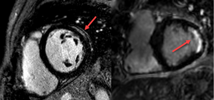
35-year-old patient with unexplained chest pain. The late recordings after the administration of contrast agent show a persistent accumulation in the outer part of the heart muscle (light area, red arrow), corresponding to a circumscribed scarring as a result of an inflammation of the myocardium. On the other hand, the remaining parts of the heart muscle remain black and are therefore not affected. Right: 70-year-old patient with coronary artery disease and bypass surgery. Here, the late images after contrast medium also show a persistent enrichment, but in the inner part of the heart muscle (light area, red arrow). This correponds to scars after a heart attack. It can be seen on the image, that the scar does not extend to the outermost parts of the heart muscle, so the scar is not transmural.
Please note that it is not possible to perform a cardiac examination using an MRI in Schlieren with a pacemaker or an implanted defibrillator. Likewise, the patients should not suffer from claustrophobia. If you have questions please call us.
Due to the variety of modalities, the choice of the optimal imaging for the individual patient is not always easy. Please do not hesitate to contact us directly if you have any questions or concerns (direct medical hotline via +41 44 255 15 01).