Bone healing is multifactorial and depends on:
- Intact blood supply
- Vital bone cells: bone formation
- Sufficient stability following fractures
Definition and example of pseudarthrosis
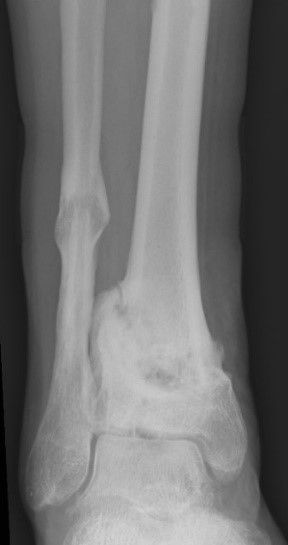
X-ray of an open fracture
The x-rays show a break as the expression of an unhealed bone fracture = pseudarthrosis (formation of false joint) just above the upper ankle joint.
Case study
In general, there is an increased risk of developing pseudarthrosis following an open fracture.
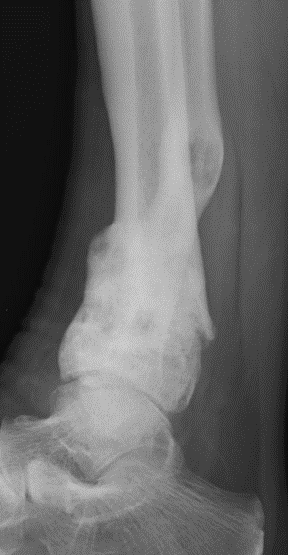
X-ray of an open fracture
The following injuries cause interruptions to the blood supply:
- Open fracture
- Severe soft tissue damage
Unstable osteosynthesis – a risk factor for the development of pseudarthrosis
Case studies
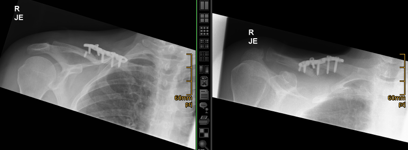
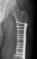
Plate broken and pulled away from the clavicle
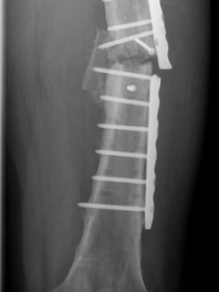
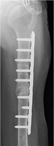
Broken plate in the thigh
Broken plate in the lower leg
X-ray diagnostics
Case study
Conventional x-rays do not always provide clear information facilitating the diagnosis of pseudarthrosis!
X-ray images of the lower leg. Has the bone healed?
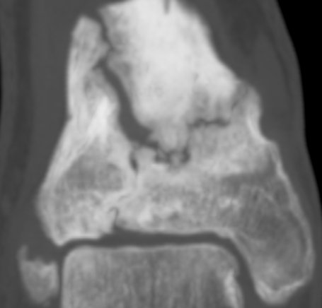
Additional diagnostic procedures with computed tomography images of the pseudarthrosis region show findings typical of pseudarthrosis
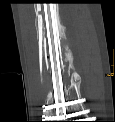
The CT images show a broken screw and a gap in the bone
The following therapeutic principles are applied when treating pseudarthrosis
- Increased stability:
- nail filling the medullary cavity
- fixed-angle bridge plate
- Correction of bone malpositioning
- Biological activation
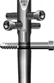
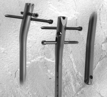
Implants to stabilise the bone: nail filling the medullary cavity
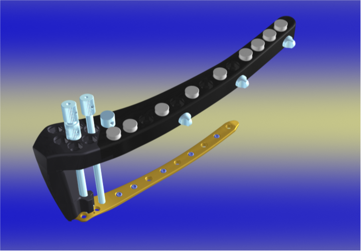
Implants to stabilise the bone: modern fixed-angle bridge plate
Case studies of the surgical treatment of pseudarthrosis
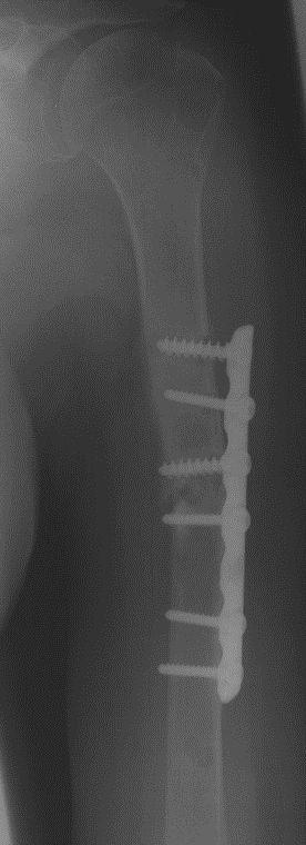
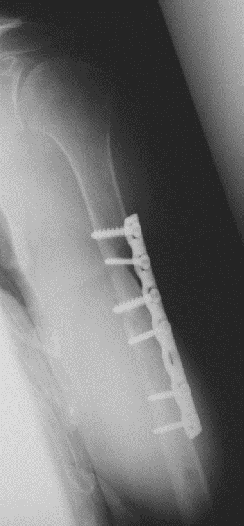
- X-ray analysis of humeral shaft pseudarthrosis following plate osteosynthesis shows the following:
- The plate and screws have become loose
- The fracture has not healed
- The bone is unstable
Operation:
- Removal of the loose plate
- Resection of the scar tissue which is preventing the formation of new bone
- Substitution of long fixed-angle bridging plate
- Grafting of autologous bone and application of bone growth factor BMP-2
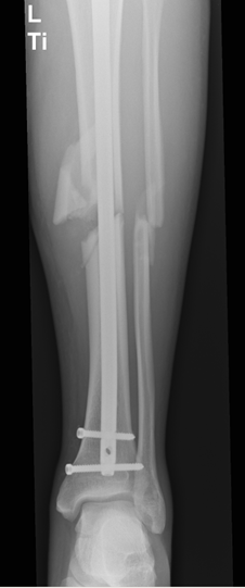
- X-ray analysis of tibial pseudarthrosis following insertion of intramedullary nail shows the following
- The fracture has not healed
- The intramedullary nail is unstable and the distal locking screw has been removed
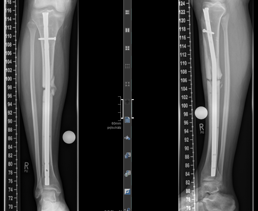
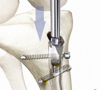
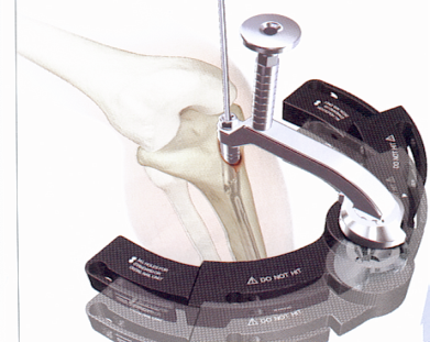
The standard surgical treatment for pseudarthrosis of the lower leg entails replacing the intramedullary nail:
- Reaming and thus “refreshing” the bone marrow cavity
- Implantation of a stable intramedullary nail
- Compression of the pseudarthrosis zone
- Stability increased by inserting locking screws in distal region
- The image shows complete bone regeneration in the former pseudarthrosis zone after a period of 3 months
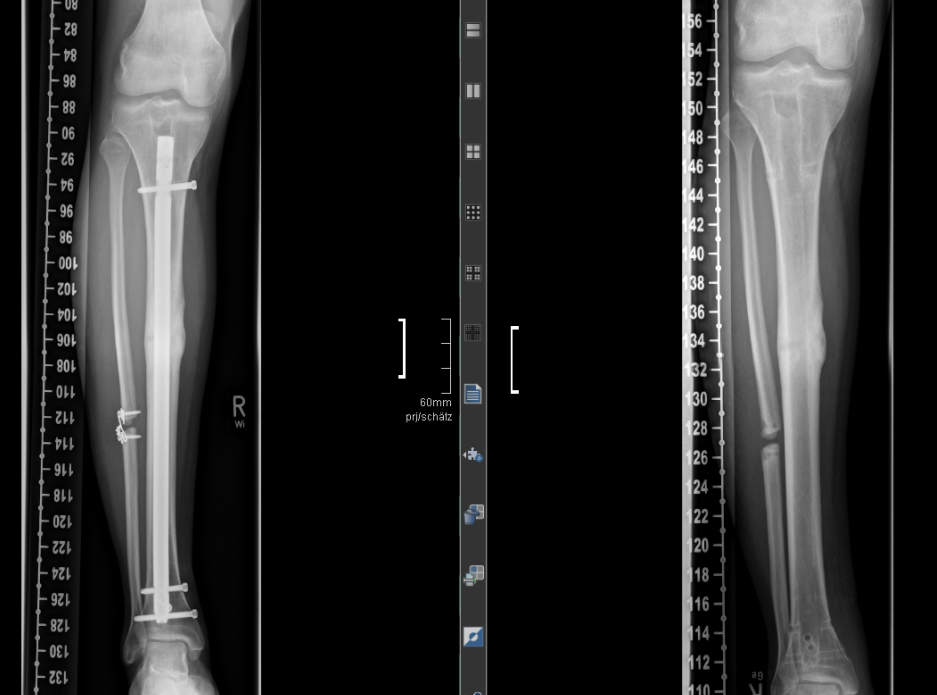
The image on the right shows the removal of the metal after approx. 1 year
The following procedures are used for the biological activation of the pseudarthrosis region
- Grafting of bone from the iliac crest
- Application of bone growth factors
- Blood collection from the iliac crest
The RIA system is another way of harvesting autologous bone
- Harvesting cancellous bone from the osseous canal, e.g. in the thigh