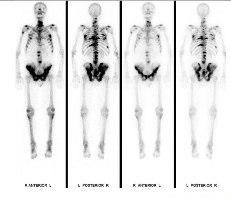
Figure: Bone scintigraphy in a patient with prostate cancer.
Skeletal scintigraphy is used in the search for skeletal metastases, in rheumatologic diseases, in the search for inflammation and infection, and in suspected child abuse.
Figure: Bone scintigraphy in a patient with prostate cancer.
At the beginning of the examination, we will inject a bone-seeking, slightly radioactive substance into one of your arm veins. In this way, we can examine your bone metabolism. Images are taken after 3 hours and require 30 – 60 minutes. During the time between injection and imaging, you may leave our service. There are essentially no dietary restrictions during this waiting time. It is mandatory, that you drink at least 1 – 2 liters of watery liquids (no milk products). In certain situations, we will need to take early scans right after injection, which will show us the perfusion of your soft tissues. These scans will take around 30 minutes.
After the exam no precautions are required.
The dose of ionizing radiation given for this examination is comparable to the yearly natural radiation dose in Switzerland. This dose is not affected by the number of images taken. If you know or think, that you are pregnant, please call us and let us know until noon of the day prior to the examination. You should also contact us, if you take care of children younger than 6 years. Furthermore, please note, that children or young adults are not permitted to accompany you to the examination. Side effects such as allergies are exceedingly rare. Please let us know if you experience any. The examination can also be done on children.
The evaluation of your examination takes some time. Hence, we cannot give you any results right after the examination. We will transmit the results and the images taken to the referring physician as soon as they are ready.
Senior Attending Physician, Vice Director of the Institute, Department of Nuclear Medicine
Attending Physician, Department of Nuclear Medicine
Attending Physician with extended responsibilites, Department of Nuclear Medicine
Attending Physician, Department of Nuclear Medicine
Attending Physician with extended responsibilites, Department of Nuclear Medicine
As a patient, you cannot register directly for a consultation. Please have your family doctor, specialist refer you. If you have any questions, please use our contact form.
Simply assign your patient via registration form.
Medical information hotline: 08.00-18.00 o’clock: +41 44 255 15 03